A lot of people have one or more types of cardiovascular disease (CVD), including hypertension, coronary artery disease (CAD), heart failure (HF), stroke, and congenital cardiovascular defects.
Because of the prevalence of CAD, we will talk about the
- Anatomy of the heart.
- Physiology of the heart.
- Causes of coronary artery disease.
- Symptoms of coronary artery disease.
- Diagnosis of coronary artery disease.
- Treatment of coronary artery disease.
Anatomic and physiologic overview of the heart
The heart is a hollow, muscular organ located in the center of the thorax, where it occupies the space between the lungs and rests on the diaphragm.
The heart weight approximately 300g; the weight and size of the heart influenced by age, gender, body weight, the extent of physical exercise and heart disease.
The major heart function is to pump oxygenated blood and nutrients to the body.
The heart has 3 layers. The exterior layer of the heart is called epicardium.
The middle layer of the heart is called myocardium, is made up of muscles and fibers and is responsible for pumping action.
The inner layer, or endocardium, consists of endothelial tissue and lines the inside of the heart and valves.
The heart consists of four chambers, two top chambers (atria) and two bottom chambers (ventricles) these chambers pump blood in a very effective way in order to keep us a life.
The pumping action of the heart is accomplished by relaxation and contraction of the muscular walls of its four chambers.
During the relaxation phase, called diastole, all four chambers relax together, which allows the ventricles to fill in preparation for contraction. Diastole is routinely referred to as the period of ventricular fillings.
During the contraction phase, called systole, atrial and ventricle systole are not simultaneous events.
Atrial systole occurs first, followed by ventricular systole. This synchronization allows the ventricles to completely fill, this behavior called atrial kick, prior to ejection of blood from there chambers.
The right side of the heart made up of the right atrium and right ventricle.
The right atrium received deoxygenated blood from two large vessels.
The vessel coming from the above draining the top part of the body is superior vena cava and the vessel draining the bottom part of the body is inferior vena cava.
The right atrium pumps the blood to the right ventricle through a valve is called the tricuspid valve.
The right ventricle distributes and pumps the deoxygenated blood (venous blood) to the lung via the pulmonary artery to pulmonary circulation for oxygenation through a valve is called pulmonic valve.
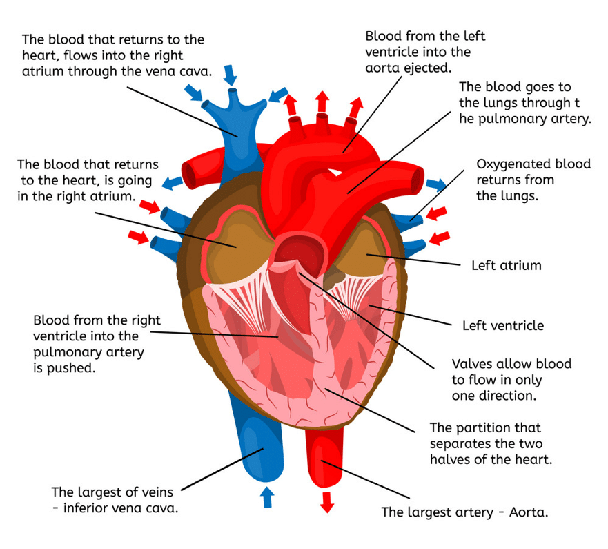
The Valves are open and close to move the blood and change the pressure within the chambers.
The left side of the heart, composed of the left atrium and left ventricle.
The left atrium received oxygenated blood from pulmonary veins and pumps it to the left ventricle through the mitral valve.
The Left ventricle distributes and pumps oxygenated blood to the body via the aorta (systemic circulation).
Two types of valves
- Atrioventricular valves: during diastole these valves are open, allowing blood to move freely from contracted atria to relaxed ventricles. These valves located between the atrium and ventricle, the right valve is called tricuspid (three leaflets) valve and the left side is called the bicuspid (two leaflets) or mitral valve.
- Semilunar valves: during systole these valves are forced to open, allowing blood to move from contracted ventricles to pulmonic and systematic circulation.
We don’t want the valve to over-expand.
So, connecting to the valves there are tendons these are called tendinous cords or sometimes referred to Latina name chordae tendineae, these days we name them tendinous cords.
And what there are is tissue which holds the valves name into the wall of the heart tissue, which holds the valves and stops them flopping back away to insure one-way flow from the atrium to the ventricle.
Tendinous cords are rooted into the wall of the heart by special muscles and these muscles that connect the tendinous cord to the wall of the heart are papillary muscles.
Papillary muscles preventing blood backflow and keeping the valve leaflets closed.
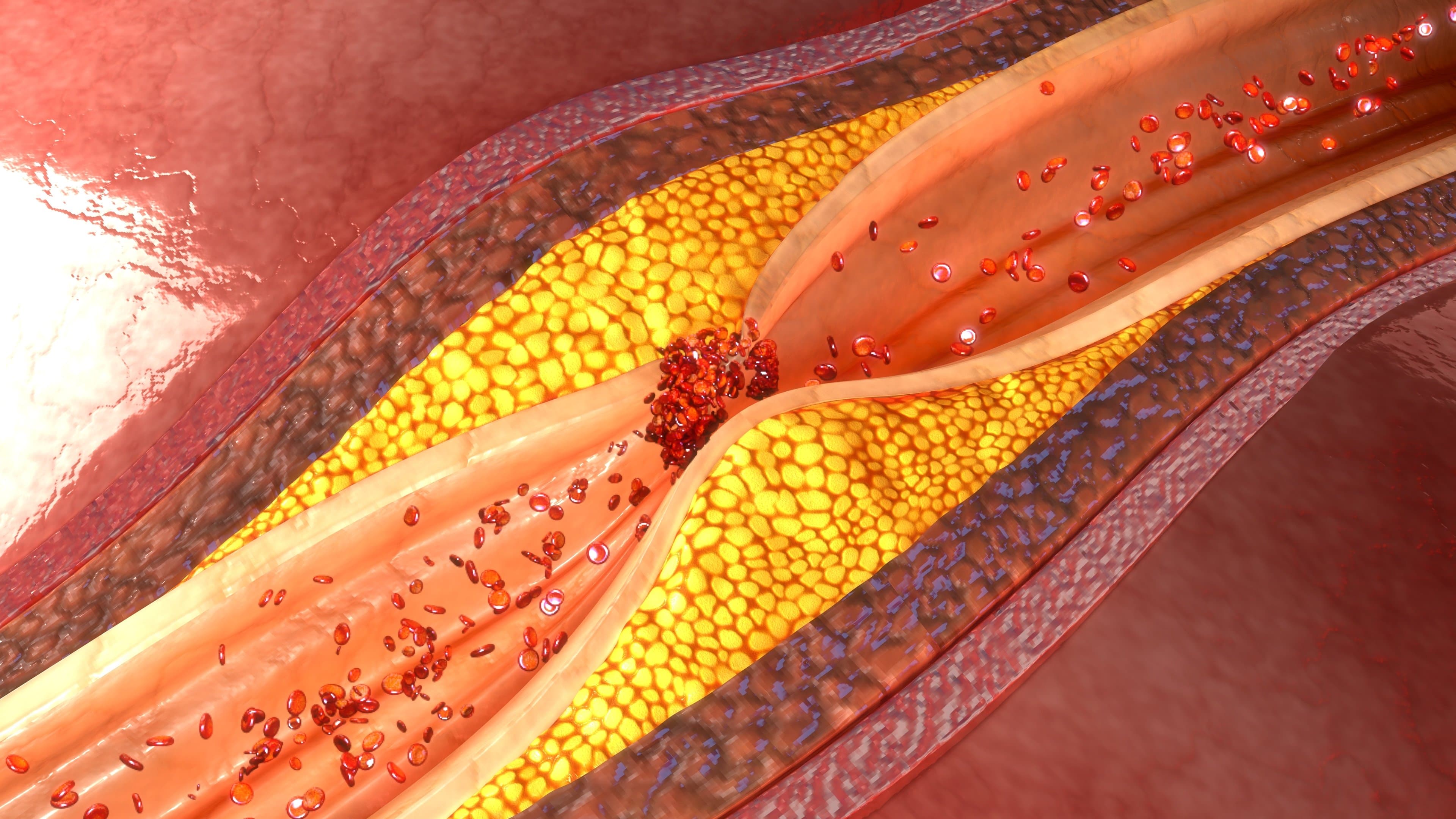
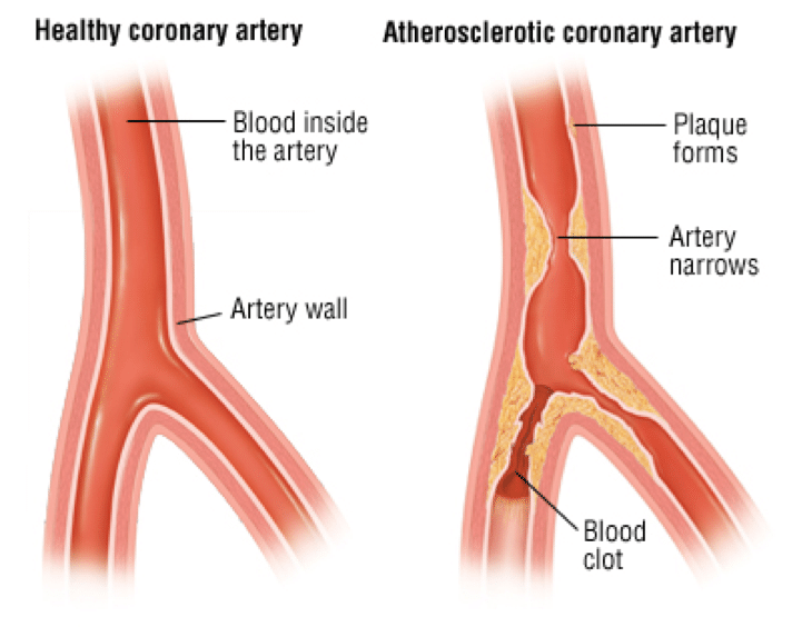
An abnormal buildup of lipid and fatty substances, and fibrous tissue in the lining of atrial blood wall.
This accumulation block and narrow the coronary vessels in a way that decreases blood flow to the heart muscles.
The development of atherosclerosis without treatment can be a life-threatening process.
Coronary Artery Disease Causes
- High blood pressure: untreated high blood pressure may result in increased stiffness of the vessel walls, leading to vessel injury. Hypertension increased the left ventricle workload cause the heart to enlarge and thicken
- Smoking: hemoglobin fuse with carbon monoxide than with oxygen. A reduced amount of oxygen may decrease the heart ability to pump
- Diabetes mellitus: high blood glucose level increase platelet formation and alter red blood cell function
- High cholesterol level: fats, lipoprotein, and density the lipid metabolism is an important contributor to the development of plaque formation
- Stress: un-coping stress may lead to hypertension and increase the potential of heart workload
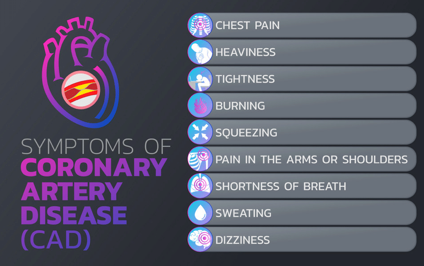
Coronary Artery Disease Symptoms
- Chest pain
- Fatigue
- Shortness of breath
- Pain in the shoulder or arm
- Sweating
- Sudden death
Risk factors
- smoke
- age
- sex
- gender
- high blood pressure
- high cholesterol level
- obesity
- physical inactivity
- diabetes
- stress
- family history
Coronary Artery Disease Diagnosis
Diagnostic tests will help your doctor to better understand and evaluate your heart disease
- Electrocardiogram to evaluate your coronary artery function during rest via electrical wave if there is an old heart attack or a new one
- Stress test to evaluate the electrical activity of your heart during exercise
- Blood tests such as cardiac marker CPK, CKMB, and T-Test. C reactive protein, lipid profile and HBA1C
- Cardiac catheterization to evaluate your coronary blood flow wither they are open or blocked
- Echocardiogram mainly to evaluate your heart muscle and left ventricle function
- Coronary imaging to evaluate your coronary blood flow
Drug therapy
- Aspirin your doctor may prescribe aspirin as a blood thinner medication to prevent blood clot formation
- Calcium channel blockers (CCB) your doctor may prescribe calcium channel blockers to improve the symptoms of chest pain
- Beta-blockers your doctor may prescribe beta-blockers to reduce your heart demand for oxygen
- Ranolazine your doctor may prescribe ranolazine to relief your chest pain
- Nitroglycerin your doctor may prescribe nitroglycerin to control your chest pain
- Angiotensin-converting enzyme (ACE) inhibitors and angiotensin ll receptor blockers your doctor may prescribe them to prevent any new episode of heart attacks
Procedure therapy
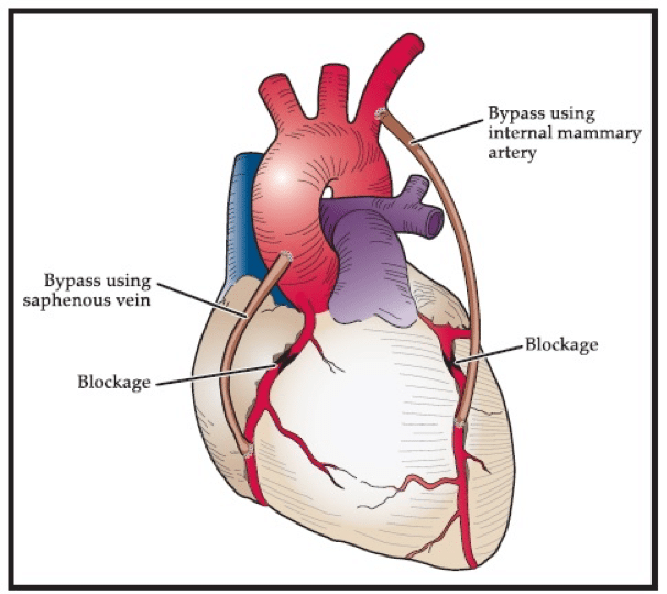
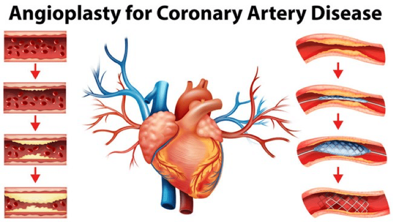
- Coronary artery bypass surgery is a surgical procedure to restore normal blood flow to an obstructed coronary artery
- Percutaneous coronary revascularization is a non-surgical procedure that use a catheter to place a stent and open up the blood vessels in the heart that have been narrowed by atherosclerosis
For more information
https://my.clevelandclinic.org/health/diseases/16898-coronary-artery-disease
https://www.webmd.com/heart-disease/guide/heart-disease-coronary-artery-disease